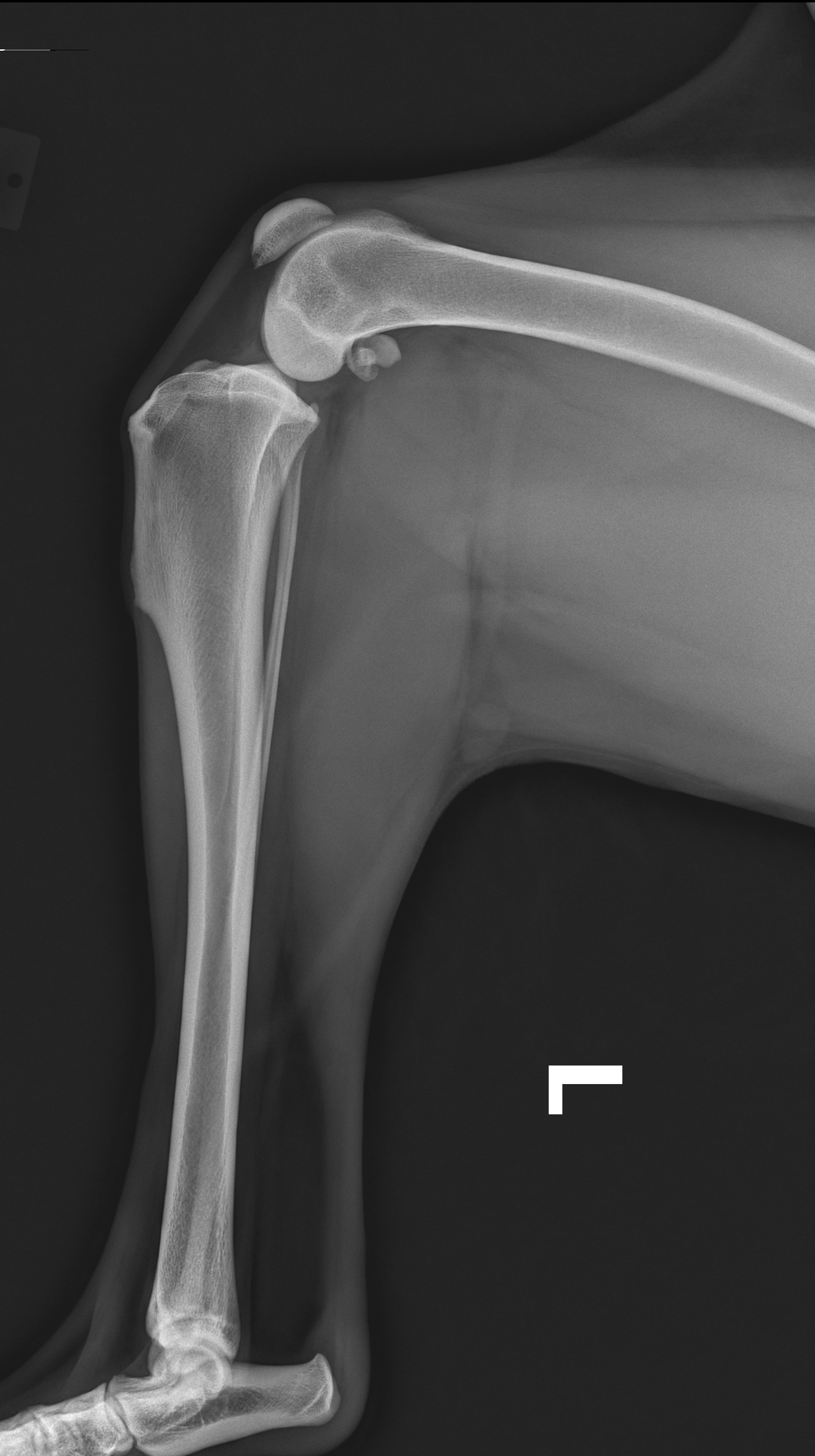
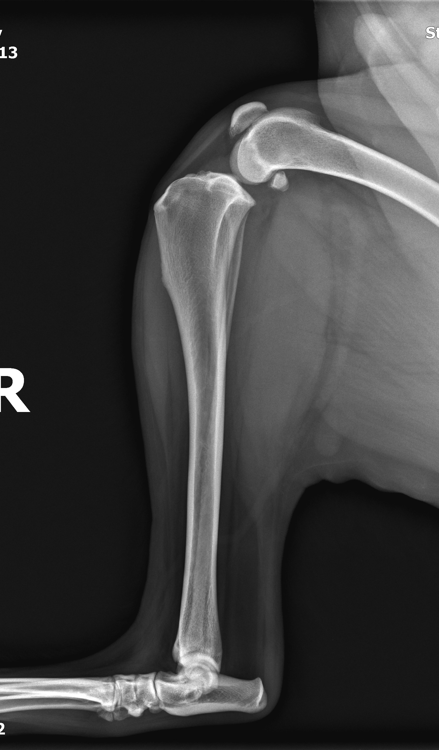
TPLO
When obtaining a stifle radiograph for TPLO (or potential TPLO), positioning is very important. Having the correct technique, positioning, and angulation are necessary for accurate measurement of the tibial plateau preoperatively.
This radiograph should be obtained prior to the day of surgery, as it reduces anesthetic time by approximately 30 minutes.
It is often best to obtain the radiograph at the time diagnosis is made, as evidence of effusion within the stifle is supportive of a CCL rupture diagnosis. It also allows assessment for other orthopedic conditions.
The proper radiograph may be obtained without sedation, depending on the patient’s temperament and cooperation, but sedation may be needed in some patients.
The tibial plateau is measured on a flexed lateral radiographic view
- Both the stifle and the tarsus must be included in the radiograph
- The stifle and the tarsus should both be at 90 degree (right) angles (90:90 view)
- Only one leg should be included in the radiographs, and the light field should be collimated appropriately
- The tarsus may need to be slightly lifted off the table to maintain stifle and hock in the same plane, allowing a true lateral (non-oblique) view of the stifle
- A perfectly lateral flexed stifle radiograph is evidenced by overlapping of the femoral condyles
For more information about TPLO and obtaining appropriate radiographs:
http://veterinarymedicine.dvm360.com/understanding-tibial-plateau-leveling-osteotomies-dogs?id=&sk=&date=&pageID=2
Examples of acceptable preoperative TPLO lateral radiographs